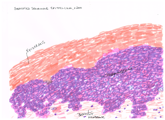
Part 1: Tissue Identification and Characterization
EPITHELIAL TISSUE
We know we are looking at epithelial tissue because of the presence of a basement membrane, and because the cells are structured as sheets of cells that line various surfaces and cavities. The cells have a "stacked" look and are joined together with cell junctions.
CONNECTIVE TISSUE
We know we are looking at connective tissue because it contains few living cells. These cells exist in a matrix, and the presence of this ground substance helps us identify tissue as connective tissue as well.
MUSCLE TISSUE
We know that we are viewing muscle tissue because the cells, or fibers, are tightly packed together, and they are aligned roughly parallel to each other.
NERVOUS TISSUE
We know we are looking at nervous tissue by the presence of two highly specialized cells, neurons and glial cells. Neurons in particular have an easily distinguished anatomy with an axon and dendrites branching out from the cell body.
Part 2:Tissue Membranes
 |
| Synovial Membrane |
Part 3: Body Position and Directional Planes
1. The blister is on her heel.
2. Dividing into front and back halves=Frontal plane
Into Left and Right halves=Midsagittal plane
Into top and bottom halves=Transverse Plane
(Sorry to write it out but I am finding it quite difficult to put on direct labels).
Part 4:Skin Structure and Function
When a single coin (penny) was placed on a subject's forearm, she experienced the light pressure sensation for 23.33 seconds. When placed on a second location on the forearm, the subject experienced the pressure for 17.95 seconds. When three more coins were placed on top of this coin, the sensation of pressured returned for 22.28 seconds. In all cases, it is likely that Meissners corpuscles are doing the majority of sensing, as Pacinian corpuscles tend to respond to rapid pressure and vibration as opposed to sustained pressure, as in the case of this experiment.


